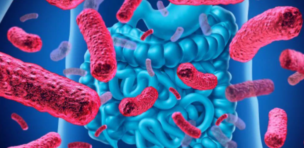
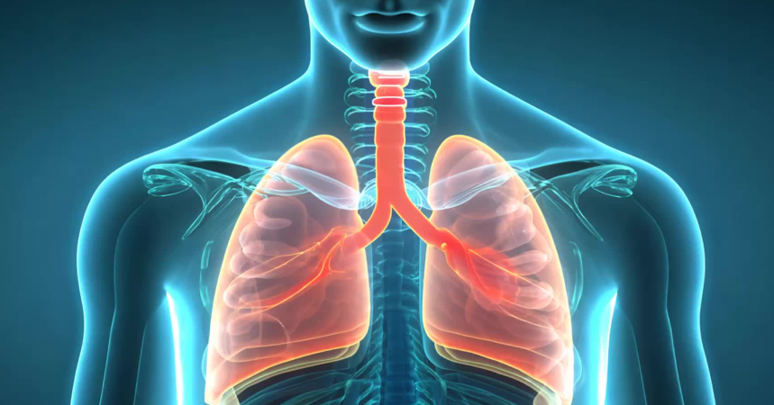
Traditionally, the intestine is primarily considered a digestive organ, while the lungs are primarily responsible for gas exchange and respiratory function. However, emerging evidence suggests the existence of a complex bidirectional communication network between these systems, namely the gut lung axis, which may significantly affect the function and health of the gut and respiratory systems. Therefore, gut microbiota not only plays an important role in metabolic processes and immune system regulation, but may also affect respiratory health.
The microbial community in the gut coexists with the host, utilizing available nutrients to perform critical tasks such as fermenting food components to produce various metabolites. For example, short chain fatty acids such as butyric acid produced by fermentation are the energy source for colon epithelial cells. Intestinal bacteria also affect liver fat metabolism, indirectly affecting cholesterol and fatty acid conversion, while promoting the synthesis of essential B vitamins and vitamin K. In addition to these metabolic processes, gut microbiota is also an essential part of immune function, as they can promote the growth and maturation of immune cells, provide local and systemic signals, and help shape immune responses. By competing for habitats and nutrients, as well as producing bacteriocins, beneficial microorganisms can prevent the reproduction of pathogenic bacteria, thereby helping to maintain tissue homeostasis.
So, how do changes in gut microbiota affect respiratory health through the gut lung axis? Dysbiosis of gut microbiota may disrupt the gut lung axis and lead to the occurrence or exacerbation of respiratory diseases such as asthma, chronic obstructive pulmonary disease, and pulmonary infections. Intestinal derived metabolites and immune signaling molecules may be involved in pulmonary inflammation and immune response.
The mechanism of action of the gut lung axis
Immune system interactions
In recent years, research on gut microbiota has been very in-depth, while research on lung microbiota is relatively scarce. For decades, the lungs were considered sterile, until the development of modern technology gradually changed this view. Research has shown that there is an interaction between the intestine and lungs that can affect the composition of the microbiota and immune response. The most striking result when comparing gut and lung microbiota is the significant difference in microbial abundance. It is estimated that the gut microbiota consists of over 100 trillion microorganisms, the vast majority of which are bacteria, with the colon being the most densely colonized part, with an estimated density of 1011 to 1012 bacteria per milliliter of content. Meanwhile, the lung microbiota is much smaller.
The composition of gut microbiota is influenced by various factors in early life, such as delivery type, feeding method, weaning time, as well as certain factors that become increasingly important with age, including body mass index, exercise frequency, lifestyle, dietary habits, and medication taken. Therefore, although the composition of gut microbiota remains largely stable, it is influenced by individual and intra individual differences throughout a person’s lifetime, thereby affecting communication within the gut lung axis. Various intestinal diseases, such as inflammatory bowel disease, ulcerative colitis, and Crohn’s disease, are associated with respiratory diseases such as asthma, chronic obstructive pulmonary disease, and cystic fibrosis.
Both gut and lung microbiota are influenced by factors such as antibiotic use, stress, diet, and metabolic diseases. The balance between the lungs and intestines depends on the integrity of the epithelial barrier, the presence of stable and diverse microbiota, the efficiency of macrophages, and the regulatory properties of T lymphocytes in the intestinal lamina propria. Inflammation caused by dysbiosis of the gut microbiota can lead to apoptosis of intestinal epithelial cells, disruption of tight epithelial junctions, and ultimately increase intestinal permeability.
In this case, not only is the exposure of microbial metabolites increased, but the exposure of various cytokines and chemokines also increases, which are produced by immune cells within the intestinal mucosal epithelium. These products are distributed in the circulatory system as well as in the lymphatic vessels of the mesentery and mediastinum, resulting in the influx of neutrophils and T lymphocytes into the intestine, forming lymphocyte aggregates that serve as a source of immune cell infiltration into other organs, including the lungs. The humoral factors transported by lymphatic vessels also reach the lungs, leading to activation of alveolar macrophages, promoting an inflammatory environment, and ultimately causing damage to the alveolar barrier. Microbial metabolites can also be distributed through similar pathways, affecting the function of lung epithelial cells and immune cells.
Mouse model studies have shown that lipopolysaccharides (LPS) can reach the lungs through damaged intestinal epithelial barriers, and increase the production of pro-inflammatory cytokines such as IL-1 β, IL-6, and TNF – α by regulating the toll like receptor TLR4 signaling pathway, leading to LPS induced acute lung injury. Fecal microbiota transplantation may play a beneficial role by restoring a balanced gut microbiota composition, including increasing the number of beneficial bacteria producing short chain fatty acids, inhibiting the activation of the TLR4 inflammatory signaling pathway, reducing the production of pro-inflammatory factors and oxidative stress.
Other microorganisms present in the gut microbiota, including segmented filamentous bacteria, may stimulate the occurrence of pulmonary pathological processes. Segmented filamentous bacteria were first discovered in experimental animals in the mid-1960s, and their characteristic biomarkers appear to be host specific, playing a key role in shaping immune processes, whether by stimulating the production of chemokines and antibacterial components, inducing intestinal lymphoid tissue, causing a strong increase in fecal IgA, or by strongly triggering Th17 cell differentiation.
In a mouse model, it was shown that segmental filamentous bacteria associated lung pathology requires activation of Th17 cells. Th17 cells induced by filamentous bacteria from the intestine are preferentially recruited to the lungs. In addition, in peripheral tissues, segmented filamentous bacteria selectively proliferate Th17 cells with dual functions, which can recognize both the antigen determining site of segmented filamentous bacteria and the T cell receptors of the host antigen determining site, thereby increasing the risk of autoimmunity.
Within the gut lung axis, interactions can also occur in the lung gut direction, not just in the gut lung direction. Based on a mouse model, intratracheal injection of LPS not only increases the number of neutrophils in bronchoalveolar lavage fluid, but also leads to changes in the intestine, manifested as an increase in the number of CD45+cells in the ileal mucosa, increased expression of goblet cells, and enhanced expression of epithelial cell adhesion molecules. Therefore, pulmonary inflammation may affect intestinal inflammation and the integrity of the intestinal barrier.
In the LPS induced systemic inflammatory response syndrome mouse model, the lungs are one of the most common and earliest affected organs, while the gut and ileum microbiota undergo 15.49% and 24.09% changes, respectively, while LPS stimulation has no effect on the structure of the colon microbiota. In the jejunum, the phylum Actinobacteria underwent significant changes, decreasing from 23.44% to 9.78%, while the phyla Bacteroidetes, Firmicutes, and Proteobacteria increased, while the phylum Verrucobacteria decreased. In the ileal microbiota, the phylum Proteobacteria showed the greatest changes, ranging from 0.44% to 6.37% and 45.7% at 4 and 8 hours after LPS stimulation, indicating the presence of dysbiosis. In addition, some respiratory viral infections can affect gut microbiota composition by inducing the production of pulmonary interferons.
Microbial interactions
The gut microbiota and its metabolites can assist in digestion and energy acquisition, nutrient absorption, vitamin production, regulate intestinal barrier integrity, stimulate the development of intestinal associated lymphoid tissue, and thus help regulate gastrointestinal homeostasis. The gut microbiota also supports the appropriate immune response of the entire organism, which requires local tolerance to external antigens while activating peripheral and systemic defense mechanisms. Therefore, these microorganisms not only regulate gastrointestinal function and immunity, but also affect distant organs such as the lungs, affecting lung microbiota and immune status.
The interaction between gut and lung microbiota affects the overall health of the host. The gastrointestinal tract and respiratory tract are directly connected through the oropharynx, maintaining indirect contact through lymph nodes and blood circulation, and also have mucosal tissue and physiological characteristics of the same origin. All of these aspects allow for interactions between gut and lung microbiota. Their interactions are mainly based on cross feeding processes and metabolites such as short chain fatty acids, and may also involve activation of host immune cells.
For example, Brucella and Lactobacillus are the main starch degrading bacteria in the gut microbiota. They can release oligosaccharides and monosaccharides, which can be utilized by themselves or other bacteria to produce short chain fatty acids. Short chain fatty acids, including acetic acid, propionic acid, and butyric acid, are involved in maintaining the normal function of the intestinal barrier, regulating glucose and lipid metabolism, alleviating oxidative stress and inflammation, and are one of the main regulators of intestinal and pulmonary immunity, maintaining the homeostasis of immune cells in the gut lung axis. Therefore, the gut microbiota is the main source of short chain fatty acids that affect immune cells in the lamina propria and mesenteric lymph nodes. Short chain fatty acids also have an impact on the production of bone marrow hematopoietic precursor cells to maintain lung homeostasis and alleviate potential airway inflammation. In a diet rich in dietary fiber, propionic acid produced by mice stimulates macrophages and dendritic cell progenitor cells, which can then trigger phagocytosis, but does not trigger Th2 mediated allergic airway inflammation.
Microbial interactions, as part of the gut lung axis, involve the activation of host immune cells. For example, studies have found that segmented filamentous bacteria in the gut microbiota can protect C57BL/6 mice from acute methicillin-resistant Staphylococcus aureus pneumonia by promoting Th17 type immunity in the lungs. Lack of these bacteria in the gut can lead to increased burden of methicillin-resistant Staphylococcus aureus in the lungs, increased lung inflammation, and higher mortality rates in mice. A mouse model has shown that segmented filamentous bacteria can also regulate the immune response to Aspergillus fumigatus lung infection through Th17 cells. Th17 cells activated by gut microbiota can reduce the production of inflammatory cytokines in lung tissue and protect mice from the effects of weight loss. Oral administration of Lactobacillus plantarum can upregulate the expression of macrophage induced C-type lectin and major histocompatibility complex MHC II on dendritic cells in the lungs, while increasing the frequency of activation and effector memory CD4+T cells, leading to a reduced bacterial burden in the lungs of mice infected with Mycobacterium tuberculosis.
The global COVID-19 caused by severe acute respiratory syndrome coronavirus 2 (SARS-CoV-2) has stimulated the scientific research community to deeply explore the potential relationship between the intestinal flora status and the infection mechanism of the virus. Some human studies reported that the imbalance of intestinal flora in COVID-19 infected people, such as the reduction of some beneficial bacteria (such as Fecal Bacillus, Eubacterium, Rosella, Lactobacillus) and the increase of opportunistic pathogens (such as Enterobacteriaceae, Enterococcus), was positively related to the SARS CoV-2 load, the aggravation of inflammation, and the severity of COVID-19.
Therefore, changes in gut microbiota, including diet related changes or antibiotic induced dysbiosis, not only lead to many gastrointestinal disorders such as malnutrition, inflammatory bowel disease, necrotizing enterocolitis, bacterial infections, but also cause changes in lung microbiota and immune response, exacerbating chronic respiratory diseases, acute infections, and worsening host conditions.
Metabolism and neural pathways
Different metabolic pathways play a crucial role in the interaction between the digestive and respiratory systems. These mechanisms involve a complex biochemical process network, including the exchange of metabolites, hormones, and signaling molecules between the gut and lungs. One of the essential metabolites is short chain fatty acids, which are produced by the gut microbiota through the metabolism of indigestible nutrients such as dietary fiber.
Most short chain fatty acids, such as acetic acid, propionic acid, and butyric acid, are consumed as energy by colon cells or used by intestinal epithelial cells to shape local immunity. The unmetabolized short chain fatty acids are subsequently redistributed from the liver to peripheral tissues through circulation. Short chain fatty acids have been shown to have anti-inflammatory and immunomodulatory effects, which may affect lung health and respiratory function. Short chain fatty acids can also affect the development of immune cells in the bone marrow, inhibit dendritic cell activity, affect regulatory T cell differentiation, and reduce the recruitment of neutrophils to inflammatory sites.
Another important pathway of the gut lung communication network is the neurotransmitters and neuropeptides produced by the intestinal nervous system within the intestine. These signaling molecules can affect the activity of the respiratory system through the vagus nerve, which connects the intestine and lungs and is responsible for regulating various physiological processes such as immune response, inflammation, and coordination of intestinal and lung function.
In addition, metabolites such as bile acids, lipids, and amino acids produced by the intestine can also affect lung function and immune response. Bile acids have been proven to have antibacterial properties and can regulate the function of lung immune cells.
The gut microbiota can interact with the lungs through soluble microbial components and bacterial metabolites (known as pathogen associated molecular patterns), which enter the bloodstream and affect metabolism and neural pathways. Pathogen related molecular patterns are recognized by host cells expressing pattern recognition receptors, which are widely expressed on various immune cells and epithelial cells. Upon activation, these receptors trigger signaling cascades to process invading pathogens and/or heal damaged tissues. Excessive receptor activation can disrupt immune homeostasis, leading to the continuous production of pro-inflammatory mediators, thereby increasing the risk of autoimmune and inflammatory diseases.
The impact of gut microbiota on lung health and disease
Asthma and allergies
Children form a stable gut microbiota around the age of 2, depending on factors such as delivery method, feeding method, and antibiotic use. Research has shown that one year old children are most susceptible to asthma due to dysbiosis of the gut microbiota. Early onset asthma is more common in children aged 3-6 months. The levels of the fecal microbiota metabolite 3-ketosheathing acid were reduced in these children. This reduction is due to a decrease in the number of Bacteroidetes producing this metabolite in the gut microbiota.
Factors that alter the gut microbiota of children aged 3-6 months, 1 year, and 3 years may affect the early onset of asthma, including the use of antibiotics during the perinatal period, delivery methods (vaginal or cesarean section), breastfeeding, and contact with pets, among others. Undoubtedly, breastfeeding can affect the gut environment beyond the gut microbiota, including its biochemical characteristics, and slightly impact the risk of asthma. It cannot be said that the delivery method will definitely have a negative impact on the occurrence of childhood asthma. However, the number of Bacteroidetes in children born by caesarean section is reduced, which may increase the risk of early-onset asthma through intestinal microbiota imbalance. No impact of perinatal antibiotics and dog ownership on fecal bacteria in children has been found yet.
It is possible to prevent the occurrence of asthma by using microbial therapies such as probiotics. These conclusions are based on the study of mouse models, which may alleviate airway inflammation in mice after treatment with four types of bacteria: Helicobacter, Veillonella, Bifidobacterium, and Rosella. The reason for using these bacterial genera is that research has found that infants at risk of asthma exhibit dysbiosis in their gut microbiota, resulting in a decrease in the abundance of these four bacterial genera.
When screening the gut microbiota of children under 13 years old for allergic eczema, food allergies, allergic rhinitis, and asthma, it was found that the greatest impact on the occurrence of allergic rhinitis is the reduction in the number of bifidobacteria in the gut. Factors affecting the decrease in their numbers include antibiotic use 6 months ago, cesarean section, and both parents having atopic skin disease. The increase of Escherichia coli/Shigella can also increase the incidence of allergic rhinitis in children. Both parents have atopic skin diseases and the use of antibiotics can stimulate the growth of these bacteria until the child is 6 months old. On the other hand, breastfeeding and the use of probiotics have a positive impact on the quantity of bifidobacteria, while also reducing the quantity of Escherichia coli/Shigella. Based on the obtained results, it is recommended to use probiotic products composed of Lactobacillus, Bifidobacterium, and Propionibacterium, supplemented with oligosaccharides, for treatment to reduce the risk of allergic rhinitis in children.
Chronic obstructive pulmonary disease
Chronic obstructive pulmonary disease is a chronic lung disease with multiple phenotypes and underlying mechanisms, associated with changes in gut microbiota composition. Compared with healthy individuals, patients with chronic obstructive pulmonary disease have different characteristics of gut microbiota, therefore, gut microbiota may play a role in the onset and progression of the disease. According to the latest data, up to 3 million people worldwide die from chronic obstructive pulmonary disease, with an average mortality rate of 42/100000. It is expected that the incidence rate will increase in the coming decades, especially in highly developed countries.
The mouse model of smoking induced chronic obstructive pulmonary disease confirmed the influence of the gut lung axis. Compared with control mice, smoking induced chronic obstructive pulmonary disease mice showed a decrease in the abundance of microorganisms such as erythridae, Bacteroidetes, and Ruminococcaceae, while the abundance of Trichomonas increased. Oral administration of vancomycin, ampicillin, or their combinations can significantly reduce the pathological occurrence of smoking induced chronic obstructive pulmonary disease, which is evident in histopathological images of lung tissue. Therefore, the composition of gut microbiota can effectively affect the disease status of patients with chronic obstructive pulmonary disease. The effect of antibiotics seems to be transferable, and the fecal microbiota of mice treated with these antibiotics can reverse the chronic obstructive pulmonary disease characteristics of recipient mice transplanted with smoking mouse microbiota.
A large-scale metagenomic study has provided interesting data on the relationship between chronic obstructive pulmonary disease and gut microbiota composition, with the potential association between the occurrence of chronic obstructive pulmonary disease and gut microbiota attributed to specific microbial taxa rather than the entire microbial community. Research has found that an increase in the abundance of genera such as Bacteroides, Actinobacteria, Lactobacillus, Flavonolyticus, and Streptomyces, as well as a decrease in the abundance of genera such as Trichomonas, Bacteroides, and Fecal cocci, are associated with the occurrence of chronic obstructive pulmonary disease. In addition, the gut microbiota score has a high predictive ability, higher than other risk factors such as gender, age, body mass index (BMI), and smoking.
tuberculosis
Mycobacterium tuberculosis is the cause of pulmonary tuberculosis, and studies on mice and humans have shown that the lung microbiota plays a role in resisting Mycobacterium tuberculosis infection. Compared with healthy controls, individuals infected with Mycobacterium tuberculosis have lower diversity of lung microbiota, and significant enrichment of Streptococcus and Pseudomonas genera. In addition, the presence of Pseudomonas aeruginosa is associated with an increased risk of treatment failure.
Mycobacterium tuberculosis infection can cause immune system dysfunction and lead to changes in gut microbiota. A study comparing the gut microbiota of adult tuberculosis patients with healthy controls found a decrease in Firmicutes, Proteobacteria, and Verrucobacteria, while an increase in Actinobacteria, Bacteroidetes, and Clostridium. In another study analyzing patients with newly diagnosed and recurrent pulmonary tuberculosis, the number of Bacteroidetes, Prevotella, and Mollusca decreased, while the number of Actinobacteria and Proteobacteria increased. Finally, in a group of pediatric patients, a decrease was observed in the genera Bifidobacterium, Dorsella, Bifidobacterium, Ruminococcus, and Prevotella, while an increase was observed in the genera Bacteroidetes, Proteobacteria, Enterococcus, and Prevotella.
Supplementing with lactobacilli can restore anti-tumor immunity, which depends on dendritic cells in the lungs. In mice infected with Mycobacterium tuberculosis, oral administration of mucin dependent or mucin dependent palmitoleic acid can strongly inhibit tuberculosis infection by epigenetic inhibition of tumor necrosis factor. The diversity of bacteria in the intestines of tuberculosis patients has also changed, which may be related to disease progression. The severity of Mycobacterium tuberculosis infection seems to be related to the gut microbiota.
Anti tuberculosis treatment includes antibiotics such as rifampicin, but it also targets bacteria other than Mycobacterium. Long term anti tuberculosis treatment can alter the patient’s gut microbiota, resulting in a dysbiosis that persists even after treatment is discontinued. Therefore, long-term anti tuberculosis treatment may make patients more susceptible to other diseases and infections.
Other viral and bacterial infections
- Influenza virus
Experimental studies have shown that symbiotic microorganisms regulate the production of virus specific CD4+and CD8+T cells and antibody responses after influenza virus infection, while antibiotic treatment can lead to gut microbiota dysbiosis and increase susceptibility to influenza infection.
Oral administration of lactic acid bacteria isolated from traditional Mongolian dairy products in influenza infected mice can alleviate infection symptoms through immunomodulatory effects. Feeding mice infected with influenza with Bifidobacterium longum can activate natural killer cells in the lungs and spleen, increase cytokine expression in the lungs, and reduce inflammation and death in the lower respiratory tract.
Nasal or oral administration of Lactobacillus plantarum can resist lethal doses of influenza A virus infection by regulating dendritic cell and macrophage activity, as well as increasing IL-12 and IFN – γ levels in bronchoalveolar lavage fluid. Lactobacillus paracasei can increase the recruitment of dendritic cells in lung tissue after infection with influenza A virus. Lactobacillus casei can also stimulate natural killer cells in the lungs. Lactobacillus rhamnosus M21 can reduce the inflammatory damage in the lungs of mice infected with influenza A virus and increase the levels of IFN – γ and IL-2 in lung lysate. The combined use of probiotic Lactobacillus mucosae and Bifidobacterium brevis can increase fecal butyrate levels in mice infected with avian influenza virus and reduce inflammatory infiltration in lung tissue.
- Respiratory syncytial virus
Mice infected with respiratory syncytial virus exhibit changes in microbial diversity, including an increase in Bacteroidetes and a decrease in Firmicutes. The increase in Bacteroidetes is mainly due to the increase in Bacteroidetes, while the decrease in Firmicutes abundance is related to the decrease in Bacteroidetes and Lactobacillaceae.
Supplementing with probiotics such as lactobacilli, bifidobacteria, enterococci, or lactobacilli before respiratory syncytial virus infection can alleviate symptoms and improve survival rates. Mucosal lactobacilli can inhibit the replication of respiratory syncytial virus and reduce the proportion of inflammatory cells in the blood, such as granulocytes and monocytes. A common bacterium, namely segmented filamentous bacteria, colonizes the intestine and can reprogram the respiratory microbiota to increase proliferation, complement production, and phagocytosis, thereby enhancing protection against respiratory syncytial virus and SARS-CoV-2 infections.
- Streptococcus pneumoniae
The gut lung axis also plays an important role in lung infections caused by pathogenic bacteria such as Streptococcus pneumoniae. These Gram positive bacteria are the main cause of pneumonia and other respiratory diseases, and their impact on host immunity is deeply influenced by the gut microbiota.
A recent study has demonstrated the interaction between gut microbiota and susceptibility to pneumococcal infection. In this experiment, wild-type mice were given broad-spectrum antibiotics (ampicillin, neomycin, metronidazole, and vancomycin) through drinking water, which effectively cleared their gut microbiota. Then, the mice were intranasal infected with Streptococcus pneumoniae. The results showed that mice lacking gut microbiota exhibited accelerated death after infection. Therefore, gut microbiota has a protective effect on the serious consequences of respiratory infections.
To further elucidate the mechanisms involved, researchers measured cytokine levels after infection. Mice treated with antibiotics to eliminate gut microbiota showed a significant increase in pro-inflammatory cytokines, such as interleukin-1 β, IL-6, and chemokine CXCL1. In contrast, the levels of TNF – α and anti-inflammatory IL-10 were significantly reduced within 6 hours after intranasal infection. The imbalance of this inflammatory response intensifies 48 hours after infection, and mice with cleared gut microbiota exhibit increased inflammation and significant tissue damage.
Alveolar macrophages are crucial immune cells in the lungs, and their ability to engulf Streptococcus pneumoniae is adversely affected in mice clearing their gut microbiota. Therefore, gut microbiota is essential in reducing inflammatory responses and is crucial in enhancing the functional activity of immune cells, especially in cases of lung infections.
The protective effect of gut microbiota on pneumonia caused by Streptococcus pneumoniae has prompted further exploration of related treatment strategies. For example, intranasal administration of probiotics has shown promise in regulating immune responses. The use of probiotics for treatment can increase the production of local TNF – α and IFN – γ, while reducing tissue damage. Therefore, probiotics have great potential as a promising adjuvant therapy in treating respiratory infections, restoring immune balance, and enhancing host defense mechanisms.
- Pseudomonas aeruginosa
Pseudomonas aeruginosa is an opportunistic pathogen, particularly notorious in respiratory infections, especially in immunocompromised individuals and populations with underlying lung diseases such as cystic fibrosis.
An emerging research field on the health effects of Pseudomonas aeruginosa also involves the gut lung axis. In a study focusing on this gut lung connection, researchers investigated the potential protective effect of probiotics on Pseudomonas aeruginosa infection. Probiotics were first administered to mice via tracheal instillation as a preventive measure. Afterwards, induce Pseudomonas aeruginosa infection through the same pathway and observe the role of probiotics during active infection. The study used a composite probiotic composed of Lactobacillus fermentum, Lactobacillus paracasei, and Lactobacillus corni. The results showed that administering probiotics significantly reduced the logarithmic growth rate of Pseudomonas aeruginosa, lowered inflammatory cytokines, and increased cell viability. Therefore, probiotics can play a meaningful role in regulating the inflammatory response associated with Pseudomonas aeruginosa infection.
lung cancer
Cancer is a major medical challenge and one of the leading causes of death among Chinese residents, with lung cancer being the leading cause of cancer death in both males and females. Due to the common embryonic origin, some similar physiological processes, and structural similarities between the gastrointestinal and respiratory tracts, it is believed that the existence of the gut lung axis is an important factor in the occurrence and potential treatment options of many diseases. Although the lungs and intestines are anatomically distant, these organs and their microbial communities still maintain close connections, indirectly through the lymphatic and circulatory systems, or directly through inhalation of gastroesophageal contents and swallowing saliva.
One way in which gut microbiota affects the development of lung tumors is through excessive intake of certain substances, such as high protein intake, which can lead to elevated protein levels in the colon. Many types of bacteria, including some Firmicutes and Bacteroidetes, can ferment amino acids into N-nitroso compounds, inducing host DNA alkylation and mutations. Through similar mechanisms, other gut microbiota metabolites such as reactive oxygen species, reactive nitrogen, and other substances that have been shown to have genotoxic (DNA damaging) effects and promote carcinogenesis also appear in the body. Other examples of metabolites in gut bacteria are deoxycholic acid and lithocholic acid, which are secondary bile acids produced by gut bacteria using bile acids. They can cause DNA damage and are associated with the occurrence of cancer.
The destruction of host microbiota can also regulate the host’s inflammatory response and cell cycle disruption. Indirect evidence of the association between intestinal flora and lung cancer also includes that the use of more antibiotics in the population will have a negative impact on the composition of intestinal flora, which is positively related to the incidence rate of lung cancer. The composition of gut microbiota rich in Bacillus species bacteria can increase susceptibility to lung cancer, while the composition of gut microbiota rich in Bifidobacterium and Bifidobacterium may have a protective effect. In addition, they can also reduce inflammation induced by TNF – α and LPS. TNF – α can induce epithelial mesenchymal transition, thereby promoting lung cancer metastasis. In addition, it has been reported that lung cancer patients have lower loads of Klebsiella, Escherichia coli, Escherichia coli, Bacteroides, and Escherichia coli. Therefore, they may serve as potential predictive factors for early lung cancer.
The causal relationship between gut microbiota and different types of lung cancer has also been the focus of many other studies. For example, using Mendelian random genome analysis, researchers identified 10 microorganisms with potential causal relationships with lung cancer, 10 with lung adenocarcinoma, 9 with lung squamous cell carcinoma, and 11 with small cell lung cancer. After adjustment, there is a strong causal relationship between Streptococcus pneumoniae and lung adenocarcinoma.
Carbohydrates available to intestinal flora can increase the production of short chain fatty acids. They can activate the production of ILC3, IL-22, regulatory T cells and Th2 cells in the lung through T cell receptor signals, thereby reducing inflammation and reducing the incidence rate of lung cancer.
The gut microbiota can also affect the treatment effectiveness of lung cancer. For example, oral administration of Lactobacillus acidophilus during cisplatin chemotherapy in a mouse model of lung cancer can increase the anti-tumor efficacy of cisplatin, reduce tumor size, and improve survival rate. There are also studies indicating that supplementing probiotics such as Clostridium butyricum before and/or after standard immune checkpoint blockade therapy can significantly prolong the progression free survival and overall survival of non-small cell lung cancer patients.
The gut microbiota has also been shown to significantly affect the treatment of immune checkpoint inhibitors, including those targeting the programmed cell death protein PD-1 and its ligand PD-L1. This effect is caused by altering the differentiation of regulatory T cells, which further induces changes in immune regulatory mechanisms. For example, the mucinous protein Akmansia increases the response to cancer immune checkpoint inhibitor therapy, and abnormalities in gut microbiota composition are associated with resistance to these treatments. In lung cancer patients, the improvement of anti-PD1 immune therapy response is positively correlated with the abundance of mucinous protein Ackermann’s bacteria. Similarly, Proteobacteria, Firmicutes, Bacteroidetes, and Actinobacteria also increase the response to PD-1 immunotherapy. Some studies have shown that drinking a large amount of yogurt can reduce the risk of lung cancer by 30%, therefore, prebiotics and probiotics may have important protective effects in the development of lung cancer.
A single microorganism may not be sufficient to induce or prevent lung cancer, but due to the synergistic effects of the host (such as the immune system), environment (such as dietary mutagens), or other microorganisms (amplification effects), the gut microbiota may play a carcinogenic role and have predictive significance, protective effects, and enhance or weaken the therapeutic effects of various forms of treatment.
Autoimmune diseases
Autoimmune diseases, such as rheumatoid arthritis and systemic lupus erythematosus, often involve the intestines and lungs. The gut microbiota can affect the systemic autoimmune response and potentially impact the lungs. Recent studies have shown that dysbiosis of the gut microbiota may exacerbate autoimmune reactions, leading to the development of interstitial lung disease and other diseases in patients with systemic autoimmune diseases.
The presence of commensal bacteria in the gut microbiota, such as Bacteroides fragilis, has been shown to promote local immune responses, thereby preventing autoimmunity in further tissue regions. Rheumatoid arthritis is an example of an autoimmune disease caused by abnormal composition of gut microbiota. It stimulates the formation of self reactive T lymphocytes (Th1 and Th12), which move through peripheral immune organs and stimulate B cell differentiation into plasma cells by producing pro-inflammatory cytokines (IL-17, TNF – α, IFN – γ). This can lead to the secretion of autoantibodies, which migrate along with immune cells to synovial tissue. In synovial tissue, they induce inflammation by activating macrophages, fibroblasts, and osteoclasts, leading to arthritis and psoriasis.
summary
The gut lung axis symbolizes the fascinating and complex interplay between two key systems in the human body. This bidirectional communication network plays a crucial role in regulating immune responses and maintaining respiratory health. Under the mediation of immune interactions, metabolic byproducts, and microbial communities in both organs, gut derived signaling substances such as metabolites and immune regulatory factors can reach lung tissue through systemic circulation, thereby exerting profound effects on respiratory function and disease susceptibility.
The gut microbiota can affect respiratory system function, so it also plays an important role in preventing the occurrence of many respiratory diseases:
The first is to use probiotics, prebiotics, or synbiotics to combat respiratory diseases and regulate lung microbiota. Probiotic strains can exert their effects directly after inhalation, or indirectly through their metabolites or activated immune cells in the gastrointestinal tract. Probiotic strains exert anti-inflammatory effects by activating regulatory T cells, producing anti-inflammatory cytokines, stimulating Th1 responses to allergens, and enhancing tolerogenic dendritic cells. Probiotic strains of Lactobacillus and Bifidobacterium have been shown to reduce lung inflammation. In a prospective pilot study targeting patients with cystic fibrosis, oral administration of composite probiotics (Lactobacillus acidophilus, Lactobacillus bulgaricus, Bifidobacterium bifidum, and Streptococcus thermophilus) significantly reduced the number of lung exacerbations during a 6-month period.
The second is dietary intervention. Dietary intervention plays a key role in regulating gut microbiota, which may affect lung health through the gut lung axis. One of the most complete connections between diet, gut microbiota, and lung health is through the intake of dietary fiber. Dietary fiber, especially from fruits, vegetables, whole grains, and legumes, can be fermented by gut bacteria to produce short chain fatty acids such as acetic acid, propionic acid, and butyric acid. Short chain fatty acids, especially butyric acid, can regulate immune responses by promoting the differentiation of regulatory T cells and inhibiting the production of pro-inflammatory cytokines, thereby helping to reduce inflammation. Research has shown that higher levels of short chain fatty acids in the gut are associated with better lung function, therefore, a diet rich in dietary fiber can prevent inflammatory lung disease.
Omega-3 fatty acids are mainly found in fatty fish (such as salmon), flaxseed, and walnuts, and have well proven anti-inflammatory properties. These fatty acids can reduce the production of inflammatory mediators, such as leukotrienes and prostaglandins, which are associated with intestinal and lung inflammation. The intake of omega-3 fatty acids in the diet is associated with a reduced risk of asthma occurrence and severity, which may be due to their ability to inhibit inflammatory processes in the airways. Omega-3 fatty acids can also enhance the immune response to respiratory infections by improving the function of macrophages and neutrophils, thereby preventing excessive inflammation and lung damage.
Some antioxidants, especially polyphenols, can affect the composition of gut microbiota. For example, polyphenols in green tea, grapes, and cocoa have been shown to promote the growth of beneficial bacteria such as lactobacilli and bifidobacteria. Vitamin D plays a crucial role in intestinal and lung health by regulating the immune system. This vitamin can enhance intestinal barrier function, reduce the translocation of harmful bacteria and endotoxins into the bloodstream, otherwise it may cause systemic and pulmonary inflammation. Appropriate levels of vitamin D are associated with lower risk of respiratory infections, reduced exacerbation of asthma, and improved overall lung function. Dietary sources of vitamin D include fortified foods, high-fat fish, and mushrooms, but supplementation is usually necessary, especially in areas with limited sunlight.
The third is fecal microbiota transplantation. Fecal microbiota transplantation is the process of transferring the fecal microbiota of a healthy donor into the patient’s intestine to restore the balance of the patient’s gut microbiota and achieve the goal of treating diseases, such as through nasogastric tube/nasoduodenal tube/nasojejunal tube infusion, oral capsules, enema, or colonoscopy. This method has been used to treat severe recurrent Clostridium difficile infections that cause pseudomembranous colitis, and has shown promise in patients with inflammatory bowel disease. Fecal microbiota transplantation can significantly reduce the risk of recurrent Clostridium difficile infection and alleviate ulcerative colitis in patients with inflammatory bowel disease by increasing gut microbiota diversity and altering the microenvironment (such as increasing short chain fatty acid levels). After fecal microbiota transplantation treatment, patients with ulcerative colitis also showed relief from other problems such as idiopathic thrombocytopenic purpura and multiple sclerosis. Therefore, people’s interest in fecal microbiota transplantation treatment for diseases outside the digestive system has greatly increased.
Fecal microbiota transplantation may also have potential in regulating the gut lung axis to improve respiratory health. For example, fecal microbiota transplantation through oral gavage and a high dietary fiber diet can help alleviate lung damage in mice exposed to cigarette smoke. Fecal microbiota transplantation can alleviate weight loss and alveolar destruction in mice with emphysema. In addition, compared with untreated mice, mice with emphysema treated with fecal microbiota transplantation or high dietary fiber diet had lower levels of IL-6 and IFN – γ in bronchoalveolar lavage fluid and serum, indicating that fecal microbiota transplantation and high dietary fiber diet have anti-inflammatory effects. In a mouse model, fecal microbiota transplantation can have an impact on Klebsiella pneumoniae induced pneumonia induced sepsis through the gut lung axis, and it can restore the changes in gut microbiota diversity caused by lung infection. The use of fecal microbiota transplantation alone or after antibiotic treatment can reduce animal mortality. Compared with antibiotics, fecal microbiota transplantation is more effective in improving local pathological lung injury and enhancing airway epithelial barrier function.
In summary, gut microbiota is also the helmsman of lung health. By delving into the mechanism of action of the gut lung axis, we can provide new ideas and methods for the treatment of many intestinal and respiratory diseases. Utilizing microbial therapies such as probiotics, prebiotics, or synbiotics to combat respiratory diseases, regulating gut microbiota through dietary interventions to improve lung health, and utilizing advanced technologies such as fecal microbiota transplantation to restore gut microbiota balance are all research directions worth further exploration in the future, and may provide more effective and safer strategies for the prevention and treatment of intestinal and respiratory diseases.